Normal Eye Anatomy
The eye is complex and highly organized organ that functions much like a camera in order to provide sight. The front part of the eye is composed of the cornea, a clear structure that allows light to enter the eye. It is surrounded by the sclera, the white part of the eye, which gives the eye its structure and strength. Behind the cornea is the iris, or colored part of the eye.
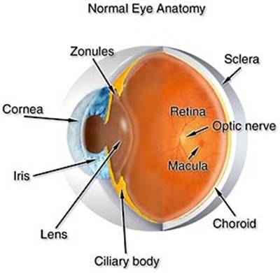

The iris gets bigger (dilates) or smaller (constricts) to regulate how much light enters the eye. Behind the iris is the lens. The lens focuses light onto the back of the eye. In some patients, the lens becomes cloudy or hazy, which is called a “cataract.” If the cataract becomes too severe and interferes with the ability of the lens to focus light, cataract surgery may be performed to remove the lens and replace it with a clear, artificial lens. Behind the lens is a clear gel-like substance called vitreous. The vitreous mainly plays an important role in the development of the eye prior to birth.
The back lining of the eye is called the retina. The retina is much like the film in a camera. It receives light and turns the light into information that is then sent to the brain through the optic nerve to provide sight. The center part of the retina is called the macula. The macula is the part that provides 20/20 vision. Its health is important for such activities as reading, driving and work-related activities.